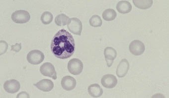
Showing posts with label HEMATOLOGY. Show all posts

Tuesday, December 30, 2014

Macrocytic Anaemias


Macrocytosis is a rise in the mean cell volume (MCV) of red cells above the normal range (in adults 80–95 fl). It is detected using a blood count, in which the MCV and other red cell indices are meas- ured. The MCV is lower in children than in adults, with a normal mean of 70 fl at 1 year of age, rising by about 1 fl each year until it reaches the adult volume at puberty.

The causes of macrocytosis fall into two groups: (i) deficiency of vitamin B12 (cobalamin) or folate (or rarely abnormalities of their metabolism), in which the bone marrow is megaloblastic (Box 2.1) and (ii) other causes (Box 2.2), in which the bone marrow is usually normoblastic. In this chapter, the two groups are considered sepa- rately. The steps to diagnose the cause of macrocytosis and subse- quently to manage it are then considered.

Iron Deficiency Anaemia


OVERVIEW

• Iron deficiency is the commonest cause of anaemia worldwide 

• Iron deficiency is usually easily diagnosed from the red cell indices

• A drop in haemoglobin is generally a late feature of iron deficiency

• The serum ferritin is a reliable means of confirming the diagnosis but may be falsely normal or even elevated as a reactive phenomenon as ferritin is an acute phase protein

• Iron deficiency is not a diagnosis in itself and in males and postmenopasual women blood loss from the gastrointestinal tract must be excluded

• Oral iron is preferred for iron replacement therapy, but occasionally parenteral iron is required


Iron deficiency is the commonest cause of anaemia worldwide and is frequently seen in general practice. Iron deficiency anaemia is caused by defective synthesis of haemoglobin, resulting in red cells that are smaller than normal (microcytic) and contain reduced amounts of haemoglobin (hypochromic).


Iron metabolism

Iron has a pivotal role in many metabolic processes, and the average adult contains 3–5 g of iron, of which two-thirds is in the oxygen- carrying molecule haemoglobin.

A normal Western diet provides about 15 mg of iron daily, of which 5–10% is absorbed (~ 1 mg), principally in the duodenum and upper jejunum, where the acidic conditions help the absorption of iron in the ferrous form. Absorption is helped by the presence of other re- ducing substances, such as hydrochloric acid and ascorbic acid. The body has the capacity to increase its iron absorption in the face of in- creased demand, for example, in pregnancy, lactation, growth spurts and iron deficiency (Box 1.1).

Once absorbed from the bowel, iron is transported across the mucosal cell to the blood, where it is carried by the protein transferrin to developing red cells in the bone marrow. Iron stores comprise fer- ritin, a labile and readily accessible source of iron and haemosiderin, an insoluble form found predominantly in macrophages.

About 1 mg of iron a day is shed from the body in urine, faeces, sweat


Haematological Findings in Health and Disease


The blood film and count in healthy individuals

The microscopic features of normal blood cells and the normal range for the blood count have been discussed in Chapter 1. In assessing what is ‘normal’ it is necessary to consider the gender, age and ethnic origin of the person being investigated.


Gender
Adult men have a higher normal range for RBC, Hb and PCV/Hct than adult women but women tend to have a somewhat higher WBC and platelet count (see Table 1.2).


Neonates, infants, and children
The blood counts of healthy neonates, infants and children differ greatly from those of healthy adults (see Table 1.3). Neonates have a higher Hb, MCV, WBC, neutrophil count and lymphocyte count than adults. Children in general have a higher lymphocyte count than adults. They tend to have a slightly lower Hb and MCV.


Pregnancy
Physiological variation in the blood count occurs during preg- nancy. The Hb falls, the MCV rises slightly and the WBC and neutrophil count rise. Immature cells (myelocytes and occasion-al promyelocytes) appear in the blood and there may be ‘toxic’granulation and Döhle bodies. 


Ethnic variation 
The blood counts of healthy Africans and Afro-Caribbeans (see Table 1.3) often show a lower white cell and neutrophil count than is usual in Caucasians (see Table 1.2). There is also a tendency to a lower platelet count, particularly in Africans. 


Abnormalities of red cells 

Polycythaemia 
Polycythaemia is an increase in the Hb. It is usually accompa- nied by an increase in the RBC and PCV/Hct. It can be caused by a true increase in the total volume of red cells in the circulation (true polycythaemia) or by a decrease in the total plasma volume (apparent or relative or pseudo-polycythaemia). It is not possible to distinguish true from apparent polycythaemia by a blood film or count. True polycythaemia is caused by overproduction of red cells. Normally red cell production is driven by erythropoietin production in response to a diminished oxygen supply to the kidney. Overproduction of red cells may be an erythropoietin- mediated physiological response to hypoxia or it may be caused by inappropriate secretion of erythropoietin or by mechanisms independent of erythropoietin. Some of the important causes of polycythaemia, classified according to mechanism, are shown in Table 4.1. 


Polycythaemia vera 

Polycythaemia vera, also referred to as polycythaemia rubra vera or primary proliferative polycythaemia, is a myeloproliferative disorder characterized by overproduction of red cells. In many patients there is also overproduction of white cells and platelets. Clinical features are facial plethora and sometimes a moderate degree of splenomegaly. The disease may be complicated by arterial thrombosis and peripheral ischaemia.
Table 4.1  Some  important causes of polycythaemia, classified according to mechanism.

Tuesday, December 23, 2014

Assessing White Cells and Platelets


White cells and platelets may be increased or decreased in number. They may also show morphological abnormalities, either inherited or acquired. Assessing whether the numbers of individual types of white cell are increased or decreased requires a differential count. However, the differential count is of little importance in itself and should only be used to calculate the absolute numbers of each cell type. The absolute counts are then compared with those expected in healthy people of the same age, sex and ethnic group. The terms used in describing numerical abnormalities in white cells and platelets are defined in Table 3.1.
Table 3.1  Terminology used  for abnormalities of white cell  and  platelet numbers

Assessing Red Cells


Red cells should be assessed as to their:
• number
• size
• shape
• degree of haemoglobinization
• distribution in the blood film.

Their appearance should be described using a standard terminology.


Assessing red cell number and distribution (anaemia, polycythaemia, rouleaux formation, red cell agglutination)

The thickness of a film of blood spread on a glass slide is deter- mined by how thick the blood is, i.e. by its viscosity. This in turn is determined by the Hb. In a normal blood film it is possible to find a part of the film which is ideal for microscopic examination where the red cells are touching but not overlapping. If the Hb is abnormally high (a condition referred to as polycythaemia) the blood has a high viscosity and the film of blood on the glass slide is thick. The red cells therefore appear packed together through- out the whole length of the film. The term ‘packed film’ is often used. Conversely, when a patient is anaemic the viscosity of the blood is low, the blood film is very thin and there are large spaces between the red cells. The effect of Hb on the blood film can be seen by comparing Figs 2.1, 2.2 and 2.3.

Usually red cells are distributed fairly regularly on the slide. Two abnormalities of distribution may occur. When there is an
Fig. 2.1  Anaemia (caused by iron  deficiency).

Monday, December 22, 2014

The Blood Film and Count (Part 2)

Haemopoietic cells
Peripheral blood cells are produced in the bone marrow. Their precursors are referred to as haemopoietic cells (Fig. 1.12). The only significant function of haemopoietic cells is the production of mature end cells. Recognizable haemopoietic precursors are present in the circulating blood of healthy subjects but, except in the neonatal period and during pregnancy, they are quite uncom- mon and are not often noted in a blood film. They are much commoner in patients with leukaemia or other haematological disorders and in patients with severe infection or other serious systemic diseases.

Myeloblasts
Myeloblasts (Fig. 1.13) are very rare in the blood of healthy subjects. They are larger than lymphocytes but often smaller than monocytes. They have a high nucleocytoplasmic ratio and scanty to moderate amounts of cytoplasm, which varies from weakly to moderately basophilic. (Basophilic in this context in- dicates a blue colour consequent on the uptake of basic dyes.) The nucleus is approximately round, nuclear chromatin is dif- fuse and nucleoli may be apparent. In patients with leukaemia and related disorders, the cytoplasm may contain small numbers of azurophilic granules or other inclusions or vacuoles (see page 91). Myeloblasts are precursors of neutrophils, eosinophils and basophils.


Promyelocytes
Promyelocytes (Fig. 1.14) are rare in the blood of healthy people. They are larger than myeloblasts with more plentiful cytoplasm and consequently a lower nucleocytoplasmic ratio. The cyto- plasm is more basophilic than that of a myeloblast and contains azurophilic (pinkish-purple) primary granules. Sometimes there is a more lightly staining zone in the cytoplasm adjacent to the nucleus, which represents the Golgi apparatus, where granules are produced. The nucleus is round or oval, is usually eccentric, shows some chromatin condensation and has a
Fig. 1.12  A diagram showing the  relationship of haemopoietic precursors to each  other and  to the  end  cells  into which they differentiate. Proliferation of cells  occurs simultaneously with maturation or differentiation so that one  myeloblast is likely to give rise  to 16 mature granulocytes and  one  proerythroblast to 16 red cells.  Myeloblasts, promyelocytes and  myelocytes are all cells capable of cell  division or mitosis. Metamyelocytes and  all later cells  are non-dividing cells.  All red cell  precursors with the exception of late  erythroblasts are dividing cells.  Myeloblasts differentiate not  only  into neutrophils, as shown in the  diagram, but also  into eosinophils and  basophils.

Friday, December 12, 2014

The Blood Film and Count (Part 1)

Blood
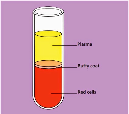
Blood is a life-sustaining fluid which circulates through the heart and blood vessels. It carries oxygen and nutrients to the tissues and waste products to the lungs, liver and kidneys, where they can be removed from the body. Usually when blood is removed from the body it forms a solid blood clot. However, if clotting is prevented by mixing with an anticoagulant, the blood separates, under the influence of gravity, into three layers (Fig. 1.1). The bottom layer is deep red in colour and is composed of red cells. The top layer is clear and pale yellow. It is called plasma and is composed of various salts and proteins dissolved in water. In between is a narrow layer called the buffy coat because of its buff or yellowish white colour. The buffy coat is composed mainly of cells of a variety of types, collectively known as white cells. In addition there are small cellular fragments, called platelets, which have a role in blood clotting.

The blood film
Although we can judge the proportions of red cells and white cells in a tube of sedimented blood, we get far more information if the blood is carefully mixed and a thin layer is spread on a glass slide to form a blood film. The blood cells are then preserved by exposure to the alcohol methanol, a process known as fixation. The fixed film of blood is stained with a mixture of several dyes so that the individual cells can be recognized when they are examined with a microscope. After staining, the colour of red 

Fig. 1.1Diagram of a tube of anticoagulated blood which has been allowed to sediment, showing the separation of blood into red cells, a buffy coat (white cells and platelets) and plasma.