
Blood
Blood is a life-sustaining fluid which circulates through the heart and blood vessels. It carries oxygen and nutrients to the tissues and waste products to the lungs, liver and kidneys, where they can be removed from the body. Usually when blood is removed from the body it forms a solid blood clot. However, if clotting is prevented by mixing with an anticoagulant, the blood separates, under the influence of gravity, into three layers (Fig. 1.1). The bottom layer is deep red in colour and is composed of red cells. The top layer is clear and pale yellow. It is called plasma and is composed of various salts and proteins dissolved in water. In between is a narrow layer called the buffy coat because of its buff or yellowish white colour. The buffy coat is composed mainly of cells of a variety of types, collectively known as white cells. In addition there are small cellular fragments, called platelets, which have a role in blood clotting.
The blood film
Although we can judge the proportions of red cells and white cells in a tube of sedimented blood, we get far more information if the blood is carefully mixed and a thin layer is spread on a glass slide to form a blood film. The blood cells are then preserved by exposure to the alcohol methanol, a process known as fixation. The fixed film of blood is stained with a mixture of several dyes so that the individual cells can be recognized when they are examined with a microscope. After staining, the colour of red
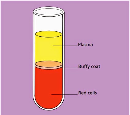 |
Fig. 1.1Diagram of a tube of anticoagulated blood which has
been allowed to sediment,
showing the separation of blood into red cells, a buffy coat (white cells and platelets) and
plasma.
|
cells is enhanced and the white cells and platelets, which would otherwise be transparent and colourless, have acquired a variety of colours which allow their detailed structure to be recognized.
One of the commonest mixtures of dyes used to stain blood cells is the May–Grünwald–Giemsa (MGG) stain, named after its inventors. All the photographs in this book are of MGG-stained blood films.
Red cells
The most numerous cells in a blood film are the red cells, also known as erythrocytes. Normal red cells are disc-shaped but are thinner in the centre (Fig. 1.2). As a consequence, on a stained blood film, they have a circular outline and a paler central area (Fig. 1.3). Red cells owe their pinkish-brown colour to the presence of a complex protein, haemoglobin, which is their major constituent. Enhancement of their colour in a stained film is because haemoglobin takes up eosin, one of the dyes of the MGG stain. In the body it is haemoglobin of the red cells which, in the
 |
| Fig. 1.2A diagram of a red cell viewed from above and in cross-section. |
 |
Fig. 1.3Normal red cells (erythrocytes) showing little
variation in size and shape, an approximately round outline and a small area of central pallor
in
some of the cells. The
small lilac-staining structures between the red cells are platelets.
|
lungs, combines with oxygen from inspired air and transports it to tissues where it is needed for the metabolic processes supplying the energy needs of the body. Mature red cells in humans (although not in some other species) differ from most body cells in that they do not have a nucleus. Red cells are produced in the bone marrow and usually lose their nuclei when they are released into the blood stream.
White cells
In healthy people there are at least five types of white cell or leucocyte in the circulating blood. Unlike red cells, white cells have retained their nuclei. The cell is therefore made up of a nucleus and cytoplasm. The cytoplasm is the site of protein synthesis and other cellular functions. The nucleus is composed of chromatin, which is mainly deoxyribonucleic
 |
| Fig. 1.4A diagram showing how white cells are classified. |
acid (DNA), carrying genetic messages. Genetic messages are transmitted from the nucleus to the cytoplasm by ribonucleic acid (RNA).
White cells are divided into granulocytes (also known as polymorphonuclear leucocytes) and mononuclear cells. There are three types of granulocyte and two types of mononuclear cell (Fig. 1.4). The names are not very logical but they have been in use for a long time and are generally accepted. Granulocytes are so named because their cytoplasm contains prominent granules.
However, monocytes also have granules and so do some lymphocytes. The term polymorphonuclear leucocyte refers to the very variable nuclear shape which is typical of granulocytes. The term mononuclear cell means that the cell has only a single nucleus. However, this is true of granulocytes, as well as of the cells conventionally referred to as mononuclear. The functions of the various leucocytes are summarized in Table 1.1.
Neutrophils
Neutrophils (Fig. 1.5) have a nucleus which stains purple and is divided into two to five segments or lobes. The lobes are separated by a thin strand or filament of nuclear material. The nuclear chromatin is heterogeneous with some clumping. The cytoplasm of neutrophils is very pale blue and is packed
with fine lilac-staining granules. The granules are referred to as
 |
Fig. 1.5A normal neutrophil with a bilobed nucleus and
cytoplasm containing
delicate lilac-staining granules. The other nucleated cell is a small lymphocyte.
|
 |
| Table 1.1The functions of leucocytes. |
neutrophilic because they owe their colour to uptake of both the acidic and the basic components of the stain. In females a proportion of the neutrophils have a very small lobe, known as a ‘drumstick’, protruding from the nucleus (Fig. 1.6). It represents the inactive X-chromosome of the cell.
Neutrophils are produced in the bone marrow. They spend 6–10 hours in the blood stream before moving from capillaries into tissues. The major function of neutrophils is as tissue phagocytes. They move preferentially to sites of infection or inflammation where they ingest, kill and break down bacteria. The process of moving to sites of infection or inflammation
 |
Fig. 1.6A normal neutrophil from a female showing a nucleus
with four lobes and a
‘drumstick’.
|
is known as chemotaxis and occurs in response to activated complement components and chemical signals released by a variety of cells. The process of ingesting bacteria is known as phagocytosis.
Eosinophils
Eosinophils (Fig. 1.7) have a nucleus that is usually bilobed and pale blue cytoplasm, which is packed with large refractile, orange–red granules. The granules are referred to as eosinophilic because they take up the acidic dye eosin. Eosinophils are produced in the bone marrow and circulate in the blood stream for
 |
Fig. 1.7A normal bilobed eosinophil. The granules are
reddish-orange and pack the cytoplasm.
|
 |
Fig. 1.8A normal basophil. The nucleus has three lobes. The
cytoplasm is packed with
large purple granules. (The lower cell is a lymphocyte.)
|
about 6 hours before migrating to tissues. They respond to chemotactic stimuli, are phagocytic and can kill ingested organisms. They are important in the body’s defences against tissue parasites, being able to discharge their granule contents extracellularly, seriously damaging large parasites. Eosinophils are also involved in allergic reactions.
Basophils
Basophils (Fig. 1.8) have a lobulated nucleus, which is often obscured by the large purple-staining granules which pack the very pale blue cytoplasm. The granules are referred to as basophilic because they take up basic components of the stain (such as methylene blue). In fact they stain metachromatically with basic stains, i.e. the granules react with a blue dye to produce a purple colour. Basophils are produced in the bone marrow and circulate in the blood in small numbers before migrating to tissue. They have a role in allergic and inflammatory responses.
Lymphocytes
Lymphocytes are the second most numerous circulating white cell after neutrophils. They are smaller than granulocytes with a round or somewhat irregular outline and pale blue, clear
 |
Fig. 1.9A large lymphocyte with a less densely staining
nucleus tha occurs in a small lymphocyte and more plentiful pale blue
cytoplasm. A nucleolus is
apparent, top left in the nucleus.
|
cytoplasm. Some lymphocytes have a variable number of azurophilic (pinkish-purple) granules. Lymphocytes are divided into three morphological categories, depending on their size, the amount of cytoplasm and the presence or absence of cytoplasmic granules. These categories are small lymphocyte (Fig. 1.5), large lymphocyte (Fig. 1.9) and large granular lymphocyte (Fig. 1.10). Small lymphocytes are most numerous. The nuclear chromatin of lymphocytes may be dense and homogeneous (particularly in small lymphocytes) or more lightly staining and somewhat heterogeneous (particularly in large lymphocytes).
 |
Fig. 1.10A large granular lymphocyte showing a moderate number
of
prominent azurophilic
granules in clear cytoplasm.
|
Occasional normal lymphocytes show a discrete but ill-defined paler structure within the nucleus, which is the nucleolus. Lymphocytes are produced from lymphoid stem cells in the bone marrow and probably the thymus. Their function is in tissues such as lymph nodes, spleen, tonsils and the lymphoid tissue associated with mucous membranes. They circulate in the blood stream, enter lymphoid tissues and emerge again from lymphoid tissues into lymphatic channels, where they form one constituent of a clear fluid known as lymph. Lymphatics drain into the thoracic duct and ultimately into the blood stream. This process of continuing movement between tissues and the blood stream is known as lymphocyte recirculation. Lymphocytes function in the body’s immune responses. They are divided into three functional types: B cells, T cells and natural killer (NK) cells. B cells differentiate in tissues into plasma cells, which secrete antibodies, thereby providing humoral immunity. T cells function in cell-mediated immunity as do NK cells. T cells also modulate B cell function. The functional categories of lymphocyte show little correlation with morphological categories except that large granular lymphocytes are either T cells or NK cells. However, other T cells cannot be distinguished morphologically from B cells. The functional categories of lymphocytes are of far more importance than the morphological categories.
Monocytes
Monocytes (Fig. 1.11) are the largest normal blood cells. They have lobulated nuclei and voluminous cytoplasm which is greyish-blue, is sometimes opaque and may be vacuolated or contai fine azurophilic granules. Monocytes have an intravascular life span of several days. They function mainly in tissues where they differentiate into long-lived macrophages (sometimes called histiocytes). Monocytes and macrophages respond to chemotactic stimuli and are phagocytic. They are part of the body’s defences against bacterial and fungal infections and also ingest and break down dead and dying body cells. They present antigen to lymphocytes.
 |
Fig. 1.11A monocyte, showing a lobulated nucleus and
voluminous, opaque cytoplasm
containing very fine azurophilic granules. Several platelets are also visible.
(Monocytes should not be confused with large granular lymphocytes. Lymphocytes have clear, pale blue
cytoplasm and discrete,
sometimes prominent granules whereas monocytes usually have more opaque, grey–blue cytoplasm with
very fine granules.)
|
They secrete chemical messengers, known as cytokines, which influence the behaviour of other body cells, including blood cells and their precursors. Monocytes differentiate not only into macrophages but also into various specialized cells, specific to different organs, such as the Kupffer cells of the liver and the microglial cells of the brain.
Platelets
Platelets are produced within the vascular channels (sinusoids) of the bone marrow by the fragmentation of the protruding cytoplasm of large bone marrow cells known as megakaryocytes. They are thus not, strictly speaking, cells but rather are fragments of the cytoplasm of cells. Platelets are considerably smaller than red cells and white cells (Fig. 1.11). They are pale blue with fine azurophilic granules which tend to be clustered in the centre of the platelet. When blood films are made, as is generally the case, from anticoagulated blood, the platelets are usually discrete and separate from each other, but in some circumstances they form clumps or aggregates.
No comments:
Post a Comment