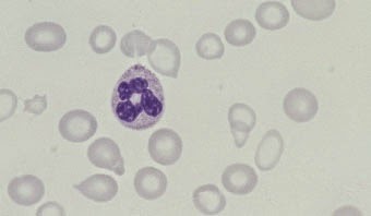
Scrapings from involved ocular site complemented with the appropriate staining method can offer the ophthalmologist circumstantial as well as definitive information concerning the identity of the invading organism.
STAINING METHODS
Light Microscopy
• KOH
• Gram’s stain
• Giemsa stain
• Grocott-Gomori Methenamine Silver (GMS) stain.
Fluorescent Microscopy
• Calcofluor white staining
• KOH-Calcofluor white procedure.
KOH mount
Principle: Potassium hydroxide is used as a mounting fluid for visualization of fungal filaments as it helps in the clearance or lyses of all surrounding tissues. In addition, Acanthamoeba cysts and Nocardia filaments can also be visualized.
Preparation of 10% KOH
• Approximately 1 gm (8 pellets) of KOH is weighed
• It is dissolved in 10 ml of distilled water
• One drop of 10% glycerol is added
• Fresh stock should be prepared every week
Can be kept at room temperature in a dropper bottle like a penicillin bottle (Fig. 4.1).
 |
| Fig. 4.1: KOH pellets and preparation of 10% KOH |