
LASIODIPLODIA THEOBROMAE
Ecology
Worldwide, well known plant pathogen.
Pathogenicity
Lasiodiplodia theobromae is a widespread saprophyte and wound-parasite on a considerable range of hosts in the tropics. It is an important parasite of bananas in storage, causing several forms of fruit-rot. Lasiodiplodia theobromae, a rare cause of mycotic keratitis and endophthalmitis.
Macroscopic Morphology
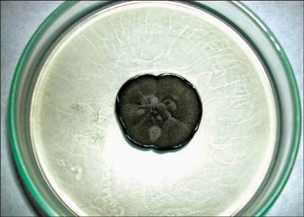
• Colonies on potato dextrose agar greyish sepia to mouse grey to black, fluffy with abundant aerial mycelium (Fig. 8.38).
• Colonies reverse fuscous black to black.
Microscopic Morphology (Fig. 8.39)
• Conidia initially unicellular, hyaline, granulose, subovoid to ellipsoide-oblong, thick- walled, base truncate; mature conidia 1-septate, cinnamon to fawn, often longitudinally striate, (18-) 20-30 × 10-15 μm.
• The pycnospores are elliptical, at first unicellular and hyaline, becoming brown and 1-septate, sometimes with longitudinal striations, 20-30 × 10-18 μm.
 |
| Fig. 8.38: Lasiodiplodia theobromae growth on potato dextrose agar (10 days) |
 |
| Fig. 8.39: Two-celled, pigmented pycnidioconidia of L. theobromae (¾Young conidia; → Matured conidia) |
NEOCOSMOSPORA VASINFECTA
Ecology
The genus Neocosmospora was established by Smith for a species apparently pathogenic to various crops including cotton, watermelon and cowpea in the southeastern United States. Neocosmospora vasinfecta, a filamentous ascomycete belonging to the Hypocreales order of the Ascomycota division is a common plant pathogen predominantly found in tropical and subtropical areas.
Pathogenicity
It has rarely been reported as being responsible for infections in humans. Known cases include a leg granuloma in a renal transplant recipient, post-traumatic osteoarthritis in an immunocompetent patient, a fatal disseminated infection in a patient with acute nonlymphocytic leukemia and an infection in a pediatric burn patient.
Occupational exposure to cotton may be a risk factor as N. vasinfecta has been isolated from intact senescent cotton roots. Although N. vasinfecta has been reported to occur in plants in India, according to our knowledge, human infections due to this fungus has not been previously described in this country. Due to their rarity, l-break/> N. vasinfecta infections are often treated as if caused by Fusarium species, because the cultural morphology.
Macroscopic Morphology
• Colonies of the fungus were fast growing on PDA at 25°C, flat, thin and appeared almost transparent at first with obvious rings forming as the colonies matured (Fig. 8.40).
• In early stage colonies are very similar to Fusarium species.
Microscopic Morphology (Figs 8.41A to C)
• Numerous ascomata (perithecia) formed within 10-14 days that gave the colony a punctate appearance (incubated at light and dark conditions at 25°C).
• Microscopic studies revealed hyaline, elongated to cylindrical conidia aggregated in slimy heads on conidiogenous cells developing on undifferentiated hyphae.
• Conidia sizes varied from 5-10 μm in length 2-3 μm wide, and were mostly single celled or with one septum.
• Some conidia appeared slightly curved. Intercalary Chlamydospores were also observed.
• The microscopic morphology was consistent with Acremonium species.
• Perithecia were orange-brown in colour, subspherical (300-400 μm in diameter), multilayered and smoothed walled, each with an apical pore.
• Cylindrical asci, 90-110-10-12 μm in diameter, were present inside the ascomata, each containing 8 ascospores in a row. The ascospores were brownish, spherical to ellipsoidal, 10-15-8-12 μm, with thick roughened walls and no germ pore present.
 |
| Fig. 8.40: Culture of Neocosmospora vasinfecta grown on potato agar at 25°C for 14 days |
 |
Figs 8.41A and B: Microscopically the perithecia are orange-brown in colour, subspherical (300-450 μm in diameter), smooth-walled and each has an apical pore |
 |
| Fig. 8.41C: Ascospores are brownish, spherical to ellipsoidal and roughed cell walls (Courtesy: Manikandan P, et al. Corneal ulcer due to Neocosmospora vasinfecta in an immunocompetent patient. Med Mycol 2007;45:Sep 18; 1-6) |
PAECILOMYCES
Pathogenicity
Paecilomyces species are rare pathogen reported into cause keratitis and endophthalmitis. They are usually present as a contaminant. Occasionally it has been reported as a pathogen in pulmonary infections, endocarditis, and sinusitis. Infections of cutaneous lesions have occasionally occurred after traumatic inoculation of the host, and Paecilomyces species have been associated with infections in patients who have had organ transplants.
Ecology
Cosmopolitan, isolated from soil and decaying plant material. Often implicated in decay of food products and cosmetics. Certain species parasitize insects.
Macroscopic Morphology (Fig. 8.42)
• Rapid growth
• Texture wooly to powdery
• Color rusty, olive brown, lilac, pinkish, beige or white on the surface, reverse pale.
Microscopic Morphology (Figs 8.43A and B)
• Hyphae septate, hyaline
• Conidiophores often branched
• Phialides thin and elongate at the tips, grouped in brush-like clusters at the ends of the conidiophores
• Conidia oval to fusoid, in long chains
• Chlamydospores sometimes present.
 |
| Fig. 8.42: Paecilomyces species growth on potato dextrose agar, 25°C, 5 days |
 |
| Figs 8.43A and B: Phialides with thinly tapered apices (→) and chains of oval conidia |
PENICILLIUM SPP
Pathogenicity
Penicillium species are usually a contaminant or a secondary invader, but infections do occur. Seven of the approximately 900 species have been isolated as etiologic agents of infection. Pulmonary infections, keratomycosis, onychomycosis, external ear infections, cutaneous lesions, bladder infections and endocarditis due to Penicililum species have been reported with, in some cases, considerable doubt about the reliability of the report.
Ecology
Cosmopolitan, predominant in regions of temperature climate. Penicillia figure among the most common types of fungi isolated from the environment. Of the approximately 150 species recognized, some are frequently implicated in the deterioration of food products, where they may elaborate mycotoxins.
Macroscopic Morphology (Figs 8.44A and B)
• Growth moderately rapid to rapid
• Texture velvety to powdery
• Color green, blue grin, grey green, white, yellow or pinkish on the surface; reverse usually pale to yellowish, rough walled red or brown.
Microscopic Appearance (Fig. 8.45)
• For species other than Penicillium marneffei, septate hyaline hyphae (1.5 to 5 μm in diameter), simple or branched conidiophores, metulae, phialides, and conidia are observed. Metulae are secondary branches that form on conidiophores. The metulae carry the flask-shaped phialides.
• The organization of the phialides at the tips of the conidiophores is very typical. They form brush-like clusters which are also referred to as “penicilli”. The conidia (2.5-5 μm in diameter) are round, unicellular, and visualized as unbranching chains at the tips of the phialides.
• In its filamentous phase, Penicillium marneffei is microscopically similar to the other Penicillium species. In its yeast phase, on the other hand, Penicillium marneffei is visualized as globose to elongated sausage-shaped cells (3 to 5 μm) that multiply by fission.
Penicillium marneffei is easily induced to produce the arthroconidial yeast-like state by subculturing the organism to an enriched medium like BHI and incubating at 35°C, in which after a week, yeast-like structures dividing by fission and hyphae with arthroconidia are formed.
 |
| Figs 8.44A and B: Penicillium species growth on potato dextrose agar, 25°C, 7 days (A—Surface; B—Reverse) |
 |
| Fig. 8.45: Penicillium species. Phialides grouped in brush-like penicilli (→) and producing conidia in chains |
PHIALOPHORA
Infections with P.verrucosa most often occur in people who work outdoors without protective clothing such as shoes and gloves. Repeated exposure and poor general health may contribute to development of the infection.
Pathogenicity
P. verrucosa can cause chromoblastomycosis or phaeohyphomycosis. The fungi are apparently introduced into host tissues through some minor injury where a contaminated splinter or thorn penetrates the skin. The infection spreads by autoinoculation and via the lymphatic system. P. verrucosa also includes several species causing diverse types of phaeohyphomycosis, presenting in the form of mycotic arthritis, subcutaneous cyst, osteomyelitis and cerebral or disseminated infection.
Ecology
Cosmopolitan, saprobes commonly isolated from decomposing wood, soil, and subaqatic debris in bodies of cold fresh water.
Macroscopic Morphology (Fig. 8.46)
• Colonies of Phialophora grow moderately slowly and attain a diameter of 2-3 cm following an incubation of 7 days at 25°C
• The texture is wooly to velvety and may be heaped and granular in some strains. From the front, the color is initially white and later becomes dark grey-green, brown or black. From the reverse, it is iron grey to black.
Microscopic Morphology (Fig. 8.47)
• Septate hyphae, phialides, and conidia are observed. The hyphae (up to 5 μm wide) are branched, and hyaline to brown. Phialophora parasitica typically produces hyphae with verrucose walls.
• In strains of Phialophora, conidial formation is Phialophora type. The phialides are usually flask- or bottle-shaped, pale brown to brown, and are terminally or laterally located on the hyphae. The length of the phialides may vary.
• The shape of the collarette varies from one Phialophora species to other. It is vase-shaped in Phialophora verrucosa, saucer- or vase-shaped in Phialophora richardsiae, and narrow with almost parallel contours in Phialophora repens and Phialophora parasitica.
• Conidia are unicellular, hyaline or brown, smooth, and round, oval or cylindrical in shape. These conidia accumulate in masses at the apices of the phialides with collarettes, giving the appearence of a vase of flowers.
 |
| Fig. 8.46: Phialophora verrucosa growth on potato dextrose agar, 25°C, 7 days |
 |
| Fig. 8.47: Phialides with vase shaped (→) collarette |
RHIZOPUS
Pathogenicity
Rhizopus is the principal agent of zygomycosis. This rapidly progressing infection is characterized by the necrosis of tissues and the production of infarcts in the brain, the lungs, and the intestines. Primarily, it is patients suffering from diabetic ketacidosis, malnutrition, severe burns, or immunocompromising conditions who are most at risk to develop this type of infection.
Ecology
Cosmopolitan, frequently isolated from soil and agricultural products (cereals, vegetables, etc). Certain species are plant pathogens.
Macroscopic Morphology (Fig. 8.48)
• Colonies of Rhizopus grow very rapidly, fill the Petri dish, and mature in 4 days.
• The texture is typically cotton-candy like. From the front, the color of the colony is white initially and turns grey to yellowish brown in time.
• The reverse is white to pale. Pathogenic species of Rhizopus can grow well at 37°C.
Microscopic Appearance (Figs 8.49A and B)
• Nonseptate or sparsely septate broad hyphae (6-15 μm in diameter), sporangiophores, rhizoids (root-like hyphae), sporangia, and sporangiospores are visualized.
• Sporangiophores are brown in color and usually unbranched.
• They can be solitary or form clusters.
• Rhizoids are located at the point where the stolons and sporangiophores meet. Sporangia (40-350 μm in diameter) are located at the tip of the sporangiophores. They are round with flattened bases.
• Apophysis is absent or rarely apparent and columellae are hemispherical. Sporangiospores (4-11 μm in diameter) are unicellular, round to ovoid in shape, hyaline to brown in color, and smooth or striated in texture.
 |
| Fig. 8.48: Rhizopus growth on potato dextrose agar, 25°C, 5 days |
 |
| Figs 8.49A and B: Rhizopus spp. sporangiophores in tufts and rhizoids (→) |
SCEDOSPORIUM APIOSPERMUM
Telemorph = Pseudallescheria boydii
Scedosporium apiospermum, previously known as Monosporium apiospermum is the anamorph of Pseudallescheria boydii, a fungus known for some time as Petriellidium boydii and Allescheria boydii.
Pathogenicity
Scedosporium apiospermum is a rare cause of endophthalmitis and keratitis. Ocular treatment of these pathologies usually proves ineffective due to the resistance of this fungus to the various antifungal agents currently marketed
An occasional agent of other infections including mycetoma, cutaneous or subcutaneous invasion, otitis, sinusitis, keratitis, endophthalmitis, pneumonia, endocarditis, meningitis, osteolyelitis, cerebral abscess and disseminated infection. Systemic infection is more commonly seen in the debilitated patient than in the normal host, but may occur in the latter under certain circumstances.
Ecology
It is a ubiquitous, earth-borne fungus commonly isolated from rural soils, from polluted water, from composts, and from manure of cattle and fowl.
Macroscopic Morphology
• Colonies of Scedosporium apiospermum grow rapidly at 25°C.
• The texture is wooly to cottony
• From the front, the color is initially white and later becomes dark gray or smoky brown. From the reverse, it is pale with brownish black zones (Figs 8.50A and B).
Microscopic Morphology (Fig. 8.51)
• Hyphae septate, hyaline
• Conidiophores with annellides simple or branced, little differentiated from vegetative hyphae
 |
Figs 8.50A and B: Scedopsorium apiospermum growth on potato dextrose agar, 25°C, 5 days (A—Surface; B—Reverse) |
 |
| Fig. 8.51: Scedosporium apiospermum conidia at the tips of more or less elongate conidophores |
• Conidia (annelloconidia) unicellular, pale brown, obovoid, with truncate bases, formed singly or in small clusters at the ends of conidiophores or from short annellidic necks arising directly from the hyphae [Scedosporium asexual state, always present (Figs 8.51 to 8.53)]
• Fascicles of conidiophores bound together in synnemata sometimes present (Graphium state)
• Brown cleistothecia often present after 2-3 weeks of incubation (sexual state Pseudallescheria boydii)
• Ascospores yellow-brown, ellipsoidal.
 |
| Figs 8.52A and B: Scedopsorium prolificans basally inflated annellides (Surface/Reverse) |
 |
| Figs 8.53A and B: Scedosporium prolificans - Basally inflated annllides (→) |
No comments:
Post a Comment