Friday, January 16, 2015

Treatment of Fungal Keratitis


MEDICAL THERAPY

The antifungal agents available today are mostly fungistatic, requiring a prolonged course of therapy. Although models of Aspergillus and Candida have been established, there are no reliable animal models of Fusarium keratitis. Fungi considered to be ocular pathogens are rarely encountered among the systemic mycoses. Thus, the therapeutic principles valid for systemic fungal infections may not apply to the cornea (O’Day DM 1987). In vitro antifungal sensitivities often are performed to assess resistance patterns of the fungal isolate. However, in vitro susceptibility testing may not correspond with in vivo clinical response because of host factors, corneal penetration of the antifungal, and difficulty in standardization of antifungal sensitivities.

Molecular-based Diagnostics for Ocular Fungal Infections


Medical laboratory technology

POLYMERASE CHAIN REACTION (PCR)


PCR is rapid diagnostic technique to detect the infectious agents even in small volume of samples. PCR is typically used for one of the following scenarios:

1. The patient presents with signs and symptoms that are most likely an infection but a definitive diagnosis cannot be made.

2. When a patient does not respond appropriately to therapy, or

3. For confirmation of a diagnosis when a patient or

4. When the patient's natural history does not coincide with his or her clinical presentation.

Tuesday, January 13, 2015

Miscellaneous Gram-Negative Bacteria


This chapter covers several Gram-negative but unrelated taxa. We did not mean to make this chapter a dumping ground, but they are all included in this chapter simply for the sake of brevity. The important taxa include the following:


• Brucella melitensis

• Bordetella pertussis

• Francisella spp.

• Pasteurella spp.

• Vibrio cholerae

• Campylobacter spp.

• Helicobacter spp.

• Legionella spp.

• Gardnerella vaginalis

• Chlamydia spp.

• Rickettsia rickettsii

Platelet Disorders


Platelets are small anucleate cells produced predominantly by the bone marrow megakaryocytes as a result of budding of the cytoplasmic membrane. Megakaryocytes are derived from the haemopoietic stem cell, which is stimulated to differentiate to mature megakaryocytes under the influence of various cytokines, including thrombopoietin. Platelets play a key role in securing primary haemostasis.
Once released from the bone marrow, young platelets are trapped in the spleen for up to 36 hours before entering the circulation, where they have a primary haemostatic role. Their normal lifespan is 7–10 days and the normal platelet count for all age groups is 150–450 × 10^9/L. The mean platelet diameter is 1–2 µm, and the normal range for cell volume (mean platelet volume; MPV) is 8–11 fL. Although platelets are non-nucleated cells, those that have recently been released from the bone marrow contain RNA and are known as reticulated platelets. They normally represent 8–16% of the total count and they indirectly indicate the state of marrow production.

Monday, January 12, 2015

The Acute Leukaemias


Acute leukaemia is a malignant disorder of white cells caused by a failure of normal differentiation of haemopoietic stem cells and pro- genitors into mature cells. This results in the accumulation of primitive leukaemic cells within the bone marrow cavity, causing bone marrow failure, and as a consequence patients typically present with anaemia, thrombocytopenia or neutropenia (Box 6.1).

Much progress has been made in understanding the pathogenesis of the acute leukaemias, and it is now clear that they occur because of the acquisition of distinct genetic abnormalities in haemopoietic stem cells or committed progenitors. These molecular abnormalities frequently occur as the result of chromosomal translocations or the loss of chromosomal material. In addition, activating mutations in genes regulating cellular proliferation, such as tyrosine kinase genes, are commonly identified. Malignant transformation of primitive cells with the capacity to develop into cells of the myeloid lineage results in acute myeloid leukaemia (AML), while acquired genetic

Thursday, January 8, 2015

Hyperkalaemia


Potassium disorders are commonly encountered in clinical practice. They are important because of the role potassium plays in determining the resting membrane potential of cells. Changes in plasma potassium mean that ‘excitable’ cells, such as nerve and muscle, may respond differently to stimuli. In the heart (which is largely muscle and nerve), the consequences can be fatal, e.g. arrhythmias.


Serum potassium and potassium balance

Serum potassium concentration is normally kept within a tight range (3.5–5.3 mmol/ L). Potassium intake is variable (30–100 mmol/day in the U K) and potassium losses (through the kidneys) usually mirror intake. The two most important factors that determine potassium excretion are the glomerular filtration rate and the plasma potassium concentration. A small amount (~5 mmol/day) is lost in the gut. Potassium balance can be disturbed if any of these fluxes is altered (Fig 11.1). An additional factor often implicated in hyperkalaemia and hypokalaemia is redistribution of potassium. Nearly all of the total body potassium (98%) is inside cells. If, for example, there is significant tissue damage, the contents of cells, including potassium, leak out into the extracellular compartment, causing potentially dangerous increases in serum potassium (see below).


Hyperkalaemia

Hyperkalaemia is one of the commonest electrolyte emergencies encountered in clinical practice. If severe (>7.0 mmol/L), it is immediately life-threatening and must be dealt with as an absolute priority; cardiac arrest may be the first mani- festation. ECG changes seen in hyperkalaemia (Fig 11.2) include the classic tall ‘tented’ T-waves and widening of the QRS complex, reflecting altered myocardial contractility. Other symptoms include muscle weakness and paraesthesiae, again reflecting involvement of nerves and muscles.

Hyperkalaemia can be categorized as due to increased intake, redistribution or decreased excretion.
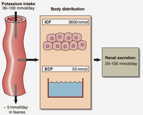
Fig 11.1 Potassium balance

Wednesday, January 7, 2015

Hypernatraemia


Hypernatraemia is an increase in the serum sodium concentration above the reference interval of 133–146 mmol/ L. Just as hyponatraemia develops because of sodium loss or water retention, so hypernatraemia develops either because of water loss or sodium gain


Water loss

Pure water loss may arise from decreased intake or excessive loss. Severe hypernatraemia due to poor intake is most often seen in elderly patients, either because they have stopped eating and drinking voluntarily, or because they are unable to get something to drink, e.g. the unconscious patient after a stroke. The failure of intake to match the ongoing insensible water loss is the cause of the hypernatraemia. Less commonly there is failure of AVP secretion or action, resulting in water loss and hypernatraemia. This is called diabetes insipidus; it is described as central if it results from failure of AVP secretion, or nephrogenic if the renal tubules do not respond to AVP. Water and sodium loss can result in hypernatraemia if the water loss exceeds the sodium loss. This can happen in osmotic diuresis, as seen in the patient with poorly controlled diabetes mellitus, or due to excessive sweating or diarrhoea, especially in children. However, loss of body fluids because of vomiting or diarrhoea usually results in hyponatraemia.