Showing posts with label BIOCHEMISTRY. Show all posts

Sunday, January 18, 2015

Intravenous fluid therapy


Does this patient need IV fluids?

The easiest and best way to give fluids is orally. The use of oral glucose and salt solutions may be life-saving in infective diarrhoea. However, patients may be unable to take fluids orally. Often the reason for this is self-evident, e.g. because the patient is comatose, or has undergone major surgery, or is vomiting. Sometimes the decision is taken to give fluids intra-venously even if the patient is able to tolerate oral fluids. This can be because there is clinical evidence of fluid depletion, or biochemical evidence of electrolyte disturbance, that is felt to be severe enough to require rapid correction (more rapid than could easily be achieved orally)

Friday, January 16, 2015

Hypokalaemia

The factors affecting potassium balance have been described previously (p. 22). Hypokalaemia may be due to reduced potassium intake, but much more frequently results from increased losses or from redistribution of potassium into cells. As with hyperkalaemia, the clinical effects of hypokalaemia are seen in ‘excitable’ tissues like nerve and muscle. Symptoms include muscle weakness, hyporeflexia and cardiac arrhythmias. Figure 12.1 shows the changes that may be found on ECG in hypokalaemia.


Diagnosis 

The cause of hypokalaemia can usually be determined from the history. Common causes include vomiting and diarrhoea, and diuretics. Where the cause is not immediately obvious, urine potassium measurement may help to guide investigations. Increased urinary potassium excretion in the face of potas- sium depletion suggests urinary loss rather than redistribution or gut loss. Equally, low or undetectable urinary potassium in this context indicates the opposite.


Reduced intake

This is a rare cause of hypokalaemia. Renal retention of potassium in response to reduced intake ensures that hypokalaemia occurs only when intake is severely restricted. Since potassium is
Fig 12.1 Typical ECG changes associated with hypokalaemia. (a) Normal ECG (lead II). (b)
Patient with hypokalaemia: note flattened T-wave. U-waves are prominent in all leads.

Thursday, January 8, 2015

Hyperkalaemia


Potassium disorders are commonly encountered in clinical practice. They are important because of the role potassium plays in determining the resting membrane potential of cells. Changes in plasma potassium mean that ‘excitable’ cells, such as nerve and muscle, may respond differently to stimuli. In the heart (which is largely muscle and nerve), the consequences can be fatal, e.g. arrhythmias.


Serum potassium and potassium balance

Serum potassium concentration is normally kept within a tight range (3.5–5.3 mmol/ L). Potassium intake is variable (30–100 mmol/day in the U K) and potassium losses (through the kidneys) usually mirror intake. The two most important factors that determine potassium excretion are the glomerular filtration rate and the plasma potassium concentration. A small amount (~5 mmol/day) is lost in the gut. Potassium balance can be disturbed if any of these fluxes is altered (Fig 11.1). An additional factor often implicated in hyperkalaemia and hypokalaemia is redistribution of potassium. Nearly all of the total body potassium (98%) is inside cells. If, for example, there is significant tissue damage, the contents of cells, including potassium, leak out into the extracellular compartment, causing potentially dangerous increases in serum potassium (see below).


Hyperkalaemia

Hyperkalaemia is one of the commonest electrolyte emergencies encountered in clinical practice. If severe (>7.0 mmol/L), it is immediately life-threatening and must be dealt with as an absolute priority; cardiac arrest may be the first mani- festation. ECG changes seen in hyperkalaemia (Fig 11.2) include the classic tall ‘tented’ T-waves and widening of the QRS complex, reflecting altered myocardial contractility. Other symptoms include muscle weakness and paraesthesiae, again reflecting involvement of nerves and muscles.

Hyperkalaemia can be categorized as due to increased intake, redistribution or decreased excretion.
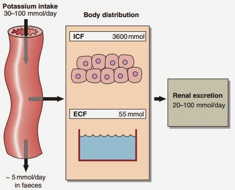
Fig 11.1 Potassium balance

Wednesday, January 7, 2015

Hypernatraemia


Hypernatraemia is an increase in the serum sodium concentration above the reference interval of 133–146 mmol/ L. Just as hyponatraemia develops because of sodium loss or water retention, so hypernatraemia develops either because of water loss or sodium gain


Water loss

Pure water loss may arise from decreased intake or excessive loss. Severe hypernatraemia due to poor intake is most often seen in elderly patients, either because they have stopped eating and drinking voluntarily, or because they are unable to get something to drink, e.g. the unconscious patient after a stroke. The failure of intake to match the ongoing insensible water loss is the cause of the hypernatraemia. Less commonly there is failure of AVP secretion or action, resulting in water loss and hypernatraemia. This is called diabetes insipidus; it is described as central if it results from failure of AVP secretion, or nephrogenic if the renal tubules do not respond to AVP. Water and sodium loss can result in hypernatraemia if the water loss exceeds the sodium loss. This can happen in osmotic diuresis, as seen in the patient with poorly controlled diabetes mellitus, or due to excessive sweating or diarrhoea, especially in children. However, loss of body fluids because of vomiting or diarrhoea usually results in hyponatraemia.

Monday, January 5, 2015

Hyponatraemia: assessment and management


Clinical assessment 

Clinicians assessing a patient with hyponatraemia should ask themselves several questions.

- Am I dealing with dangerous (life-threatening) hyponatraemia?

- Am I dealing with water retention or sodium loss?

- How should I treat this patient?

To answer these questions, they must use the patient’s history, the findings from clinical examination, and the results of laboratory investigations. Each of these may provide valuable clues.

Hyponatraemia: pathophysiology

Hyponatraemia is defined as a serum sodium concentration below the reference interval of 133–146 mmol/ L. It is the electrolyte abnormality most frequently encountered in clinical biochemistry.


Development of hyponatraemia

The serum concentration of sodium is simply a ratio, of sodium (in millimoles) to water (in litres), and hyponatraemia can arise either because of loss of sodium ions or retention of water. 

- Loss of sodium. Sodium is the main extracellular cation and plays a critical role in the maintenance of blood volume and pressure, by osmotically regulating the passive movement of water. Thus when significant sodium depletion occurs, water is lost with it, giving rise to the characteristic clinical signs associated with ECF compartment depletion. Primary sodium depletion should always be actively considered if only to be excluded; failure to do so can have fatal consequences. 

- Water retention. Retention of water in the body compartments dilutes the constituents of the extracellular space including sodium, causing hyponatraemia. Water retention occurs much more frequently than sodium loss, and where there is no evidence of fluid loss from history or examination, water retention as the mechanism becomes a near-certainty.


Water retention

The causes of hyponatraemia due to water retention are shown in Figure 8.1.
Fig 8.1 The causes of hyponatraemia

Thursday, December 25, 2014

Water and sodium balance


Body water and the electrolytes it con- tains are in a state of constant flux. We drink, we eat, we pass urine and we sweat; during all this it is important that we maintain a steady state. A motor car’s petrol tank might hold about 42 L, similar to the total body water content of the average 70 kg male. If 2 L were lost quickly from the tank it would hardly register on the fuel indicator. However, if we were to lose the same volume from our intravascular compart- ment we would be in serious trouble. We are vulnerable to changes in our fluid compartments, and a number of important homeostatic mechanisms exist to prevent or minimize these. Changes to the electrolyte concentration are also kept to a minimum. 

To survive, multicellular organisms must maintain their ECF volume. Humans deprived of fluids die after a few days from circulatory collapse as a result of the reduction in the total body water. Failure to maintain ECF volume, with the consequence of impaired blood circulation, rapidly leads to tissue death due to lack of oxygen and nutrients, and failure to remove waste products.


Water 

Normal water balance is illustrated in Figure 7.1.

Water intake largely depends on social habits and is very variable. Some people drink less than half a litre each day, and others may imbibe more than 5 L in 24 hours without harm. Thirst is rarely an overriding factor in determining intake in Western societies.
Fig 7.1 Normal water balance.